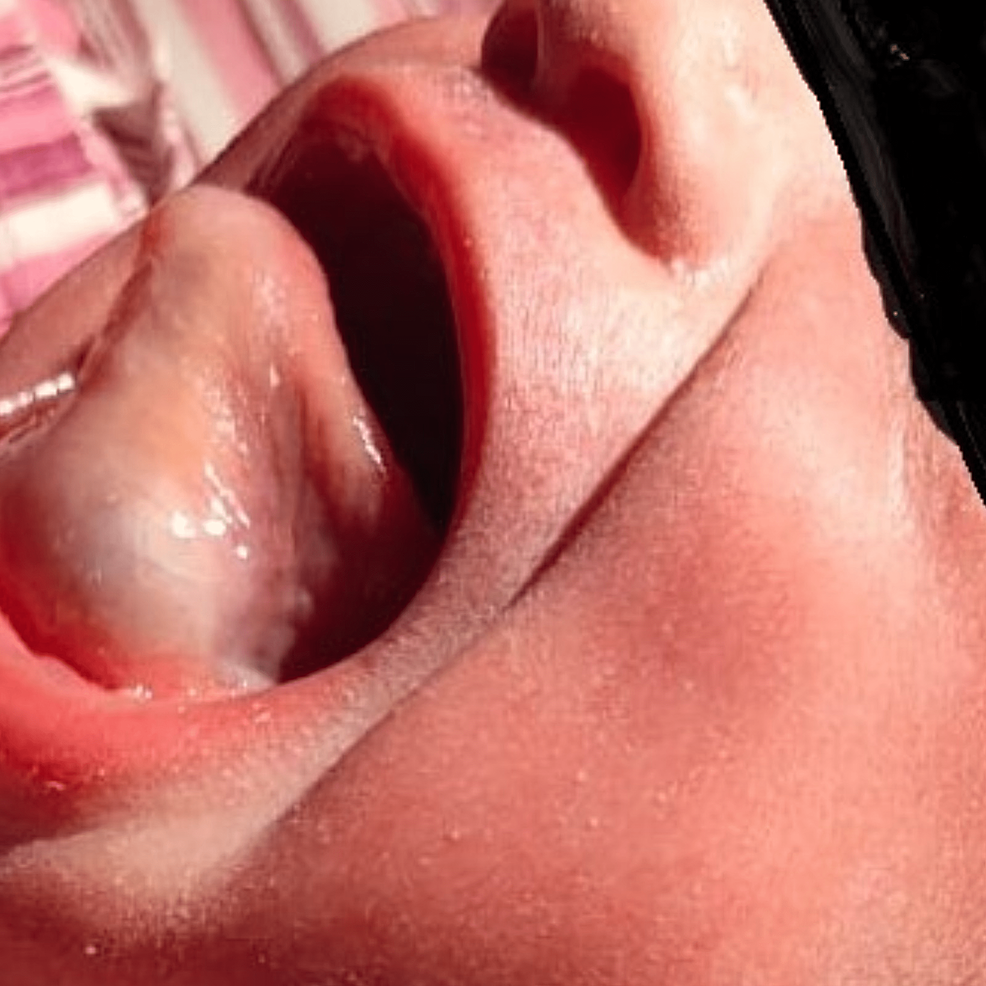
A ranula is a type of mucous cyst in the floor of the mouth. It’s often caused by an injury or blockage of the salivary glands.
Dealing with a ranula can be uncomfortable and concerning. These oral cysts, typically filled with a clear or bluish liquid, emerge from a ruptured salivary gland caused by trauma, plugging, or sometimes for no apparent reason. Generally found under the tongue, a ranula presents as a swelling that may increase in size over time but tends to be painless.
Smaller ranulas can resolve on their own, but larger or more persistent ones often require medical intervention. Treatment options range from simple drainage to more complicated surgical procedures in recurring cases. Understanding the nature of ranulas and seeking prompt treatment can help prevent potential complications such as infection or chronic discomfort.

Credit: www.cureus.com
Introduction To Ranulas
Ranulas are oral cysts that can cause discomfort and swelling beneath the tongue. These fluid-filled sacs are not uncommon and can affect both adults and children. Identified by their blue or clear mucus, ranulas are intriguing conditions within oral health. This section delves into their basics, prevalence, and differentiation from other cysts.
A ranula is a type of mucocele, or mucus cyst, that forms in the mouth. It often appears on the floor of the mouth, resulting from a blocked salivary gland. These cysts are typically painless and are filled with a thick, jelly-like substance.
- Type: Mucus retention cyst or extravasation cyst.
- Location: Floor of the mouth, near the salivary glands.
- Cause: Blocked salivary duct or trauma.
- Appearance: Clear or bluish, dome-shaped swelling.
Ranulas can occur at any age, but they are more frequent in younger individuals. They are relatively rare, with specific patterns observed in various demographics.
| Age Group | Frequency |
|---|---|
| Children and Young Adults | More Common |
| Adults | Less Common |
Ranulas have distinct features that set them apart from other oral cysts. Their unique characteristics are important for accurate diagnosis.
- Color: Often blue-tinged due to the thin tissue covering.
- Consistency: More fluid and gel-like compared to others.
- Size: Can grow larger than many other oral cysts.
Other types of oral cysts, like dermoid cysts or epidermoid cysts, typically form in other areas and have different textures. Understanding these differences aids in prompt and proper treatment.

Credit: www.nejm.org
Causes And Pathophysiology Of Ranulas
Ranulas are fluid-filled cysts that appear in the floor of the mouth. They occur when a blockage or injury affects the salivary glands. Understanding their causes and pathophysiology helps in diagnosis and treatment. Let’s dive into the specifics, starting with how they develop.
Etiology: How Ranulas Develop
Ranulas occur when salivary gland ducts get blocked, causing saliva to collect and form a cyst. These cysts often happen in the sublingual glands located below the tongue.
- Salivary gland blockage leads to ranula formation.
- Sublingual glands are primarily affected.
Anatomical Considerations And Salivary Glands
The human mouth has several salivary glands. The sublingual, submandibular, and parotid glands are the major ones. Ranulas usually form in the sublingual glands. These are under the tongue and maintain mouth moisture and help in digestion.
| Gland | Location | Function |
|---|---|---|
| Sublingual | Under the tongue | Moisturize mouth, digestion aid |
| Submandibular | Beneath the jaw | Produce saliva |
| Parotid | Near the ears | Release saliva into mouth |
The Role Of Trauma And Obstruction In Ranula Formation
Injury to the mouth floor or salivary glands can lead to ranula development. Trauma or obstruction prevents saliva from flowing normally, which results in accumulation and cyst creation.
Common sources of trauma and obstruction include:
- Lip biting or piercings can cause direct injury.
- Dental procedures may inadvertently damage ducts.
- Sialoliths, or salivary stones, are a form of obstruction.
Types Of Ranulas
Welcome to our exploration of the different forms of ranulas. These fluid-filled cysts can appear in various ways under the tongue and in the mouth. Two main types of ranulas exist, and understanding them is crucial for anyone facing this condition or just curious about it.
Simple Ranulas: Features And Implications
Simple ranulas present as clear, blueish lumps on the floor of your mouth. They resemble a small balloon filled with liquid and are usually not larger than a cherry. Simple ranulas arise from the blockage of saliva in a minor salivary gland. While they may look alarming, they’re typically pain-free and benign. Still, their presence can affect your comfort and mouth functionality. Consultation with a health care provider is advisable if you spot one.
Plunging Ranulas: Understanding Deep Extension
- A plunging ranula occurs when the cyst extends deeper into the neck.
- This type may not always be visible inside the mouth, making it trickier to detect.
- It can cause swelling, discomfort, and sometimes even impact speech and swallowing.
- Professional evaluation becomes imperative for a potential plunging ranula. An ultrasound or MRI may be necessary for a complete diagnosis.
Causes And Implications Of Diverging Ranula Types
Generally, a ranula develops when a salivary gland is injured or its duct is blocked. Simple and plunging ranulas distinguished primarily by their size and location, share common origins. These may include:
| Cause | Simple Ranula | Plunging Ranula |
|---|---|---|
| Local trauma | Yes | Often |
| Salivary gland blockage | Common | Common |
| Surgical complications | Rarely | Possible |
Despite sharing causes, the implications for simple and plunging ranulas can differ significantly. Simple ranulas often resolve with minimal treatment or even spontaneously. On the other hand, plunging ranulas might require surgical intervention. Therefore, understanding the type of ranula is essential for appropriate care and management.
Clinical Manifestations Of Ranulas
Clinical manifestations of ranulas offer insights into the presence of this mouth condition. A ranula is a type of mucocele, or cyst, that forms in the floor of the mouth. It occurs when a salivary gland is blocked. This leads to a swelling filled with fluid. Knowing the symptoms early can help people seek timely treatment.
Symptoms: Recognizing A Ranula
Ranulas typically present as:
- Soft, bluish swellings in the mouth floor
- Painlessness, making them hard to notice at first
- Size variation from small to large, affecting comfort
Some might feel a lump when speaking or eating.
Complications: When Ranulas Become Troublesome
Ranulas become complicated when:
- They increase in size, leading to discomfort
- Speech or chewing is affected
- Secondary infections occur
Infection signs include redness, pain, and pus.
Associated Conditions: Co-occurring Health Issues
Health issues linked with ranulas are:
| Condition | Explanation |
|---|---|
| Sialadenitis | Inflammation of salivary glands |
| Salivary Gland Stones | Blocks saliva flow |
Ranulas may indicate an underlying salivary gland disorder.
Diagnostic Strategies For Ranulas
Understanding Ranulas requires effective diagnostic strategies. These fluid-filled cysts, commonly found in the floor of the mouth, present unique challenges for diagnosis. To ensure accurate identification and treatment planning, thorough clinical evaluation, imaging, and sometimes fine-needle aspiration become essential tools. Let’s explore the most effective strategies to diagnose Ranulas.
Clinical Examination And History Taking
The first step in diagnosing a Ranula involves a comprehensive clinical examination. Doctors look for a blue or translucent swelling under the tongue. They consider the lump’s size, shape, and location. A patient’s history offers clues about the onset and progression of symptoms. This background is crucial for differentiating Ranulas from other oral conditions.
Imaging Techniques In Ranula Evaluation
Imaging plays a pivotal role in Ranula assessment. Ultrasound imaging is non-invasive and helps visualize the cyst’s content. Similarly, Magnetic Resonance Imaging (MRI) provides detailed pictures of the Ranula and surrounding tissues. This helps in understanding the cyst’s relation to the sublingual and submandibular glands.
- Ultrasound: Safe and quick, shows fluid-filled structures.
- MRI: Offers comprehensive details, but is more expensive and time-consuming.
Fine-needle Aspiration: Pros And Cons
Fine-needle aspiration (FNA) is a diagnostic technique where a thin needle draws fluid from the Ranula. While FNA can confirm the cystic nature of the swelling, it is not without risks or limitations.
| Pros | Cons |
|---|---|
| Minimally invasive | Potential for cyst recurrence |
| Quick and simple procedure | May not differentiate between types of cysts |
| Immediate results on cyst content | Discomfort or pain at the aspiration site |

Credit: en.wikipedia.org
Therapeutic Approaches To Ranulas
A ranula is a type of cyst that can form under the tongue. It can look swollen or cause discomfort. This blog post explores how doctors treat ranulas. Some treatments are with surgery and some are not.
Surgical Intervention: Techniques And Outcomes
When a ranula gets too big or bothers a lot, doctors may suggest removing it with surgery. The main goal is to take away the cyst and stop it from coming back.
- Marsupialization: This method involves making a cut on the ranula to let the fluid out. The edges are stitched to stop the cyst from sealing up too quickly.
- Excision: This is surgical removal of the entire ranula. It is a thorough approach and often prevents the ranula from coming back.
The success rate of these surgeries is high. Yet, there can be risks like damage to nearby nerves.
Non-surgical Management: When Is It An Option?
Not all ranulas need surgery. Small ones might go away on their own. Doctors can also drain them without making big cuts.
- Aspiration: This is when doctors use a needle to remove the fluid inside the ranula.
- Sclerotherapy: A special solution is injected to shrink the ranula.
These methods can help, especially if surgery is too risky.
Postoperative Care And Prevention Of Recurrence
After surgery, it is important to take care of the mouth area. This helps heal and stops the ranula from coming back.
| Step | Action |
|---|---|
| 1 | Keep the area clean with salt water rinses. |
| 2 | Eat soft foods to avoid hurting the surgery spot. |
| 3 | Check with the doctor before using a toothbrush near the area. |
| 4 | Go for check-ups to make sure the ranula is gone. |
Following the doctor’s advice is key for good healing and stopping ranulas from forming again.
Conclusion
Understanding ranulas is important for managing oral health. These mucus-filled cysts can be uncomfortable, but options exist for relief. Timely consultation with a healthcare professional ensures proper diagnosis and treatment. Remember, addressing oral issues early maintains overall well-being and prevents complications.
Embrace dental health; prioritize your smile today.

